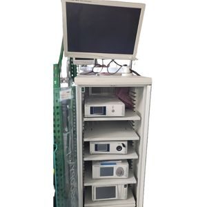
Description
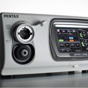
This powerful HD platform offers crisp, clear imaging, and the use of i-scan technology may help improve lesion detection and characterization. The EPK-i7000 is the first endoscopy processor to offer integrated HD recording to collect and share findings. All of these advanced features are easily accessed through an intuitive user interface. The EPK-i7000’s HD image processing is designed to provide enhanced, highly detailed live and recorded videos. This physician-controlled per-pixel modification of HD white light images enhances mucosal surface texture and visualization of blood vessels in three algorithms: SE, CE, and TE. In addition, the unique twin mode displays both the white light and enhanced i-scan image simultaneously. Physicians can switch seamlessly in real time among the three i-scan modes and white light image to view multiple aspects of tissue structure, which may increase disease detection and characterization.