ZEISS Cirrus 5000 HD OCT

Refurbished

Refurbished

ZEISS HD-OCT 5000 has high resolution visualization capabilities and sophisticated clinical applications.

Final price will be confirmed in quote

View cart above to complete quote request.

ZEISS HD-OCT 5000 has high resolution visualization capabilities and sophisticated clinical applications.

Categories: ,

Description

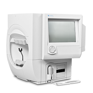
Zeiss HD-OCT 5000 has advanced RPE analysis to track retinal pigment epithelial integrity and Ganglion Cell Analysis to assess glaucomatous loss in the macula. FastTrac™ retinal tracking helps prevent eye motion artifacts. This feature allows for the highest resolution B-Scans to be captured in identical locations throughout the patient’s visit history providing precise evaluation of change in targeted pathologies.

CIRRUS HD-OCT 5000 is specifically designed to deliver a carefully constructed set of sophisticated applications that build upon one another to deliver rapidly-evolving diagnostics for multiple patient populations. CIRRUS HD-OCT 5000 is the clinical powerhouse with FastTrac, and sophisticated analyses for the busy advanced care practice.

  • HD Smart Scans
  • FastTracâ„¢ retinal tracking system
  • Macular Thickness OU Analysis
  • Advanced RPE analysis
  • Ganglion Cell Analysis
  • Guided Progression Analysis (GPAâ„¢)
  • Precision FoveaFinderâ„¢
  • Macular Thickness and Change Analysis
  • Macular Thickness Normative Data

Specifications

  • Weight: 80 lbs (36 kg)
  • Dimensions of instrument: 26L x 18W x 21H (in), 65L x 46W x 53H (cm)
  • Dimensions of table: 39L x 22W (in), 99L x 56W (cm)
  • OCT Imaging Methodology: Spectral domain OCT
  • Optical source: Superluminescent diode (SLD), 840 nm
  • Scan speed: 27K- 68K A-scans per second
  • A-scan: 2.0 mm (in tissue), 1024
  • Axial resolution: 5 μm (in tissue)
  • Transverse resolution: 15 μm (in tissue)
  • Fundus Imaging Methodology: Line scanning opthalmoscope (LSO)
  • Live fundus image: During alignment and during OCT scan
  • Optical source: Superluminescent diode (SLD), 750 nm
  • Field of view: 36 degrees W x 30 degrees H
  • Frame rate: > 20 Hz
  • Transverse resolution: 25 μm (in tissue)
  • Iris Imaging Methodology: CCD camera
  • Resolution: 1280 x 1024
  • Live iris image: During alignment
  • Fixation: Internal, external
  • Internal fixation focus adjustment: -20D to +20D (diopters)
  • Electrical rating (115V): Single Phase, 100–120V~ systems: 50/60Hz, 5A
  • Electrical rating (230V): Single Phase, 220–240V~ systems: 50/60Hz, 2.5A
Never miss out on available equipment!

Sign up to download a PDF of our current, complete inventory list.  Also, by signing up, you'll be the first to receive full access to weekly inventory updates, recently received products, and our latest featured equipment.